Prof. Hagai Abeliovich
Major research interests:
Yeast Cell Biology and Biotechnology
Current research projects:
- Understanding the mechanism of regulation of nutrient–starvation induced autophagy in yeast and its potential implication for industrial fermentations.
- Understanding the mechanism of mitochondrial turnover through mitophagy in the yeast S. cerevisiae.
- Selective effects of weak organic acid food preservatives on intracellular membrane trafficking.
Metabolic engineering of yeast for the production of plant nutraceuticals.
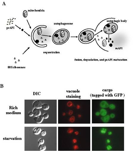
A. Overview of membrane trafficking in yeast macroautophagy.
During autophagy, various components of the cytoplasm, such as mitochondria, ribosomes, specific cargo such as prAPI (see part B of this figure), and soluble cytoplasmic material (depicted as diffuse gray) are sequestered into autophagosomes, which are large (300–900 nm) double bilayer vesicles. Once formed, autophagosomes fuse with the vacuole, releasing a single–bilayer bound autophagic body into the lumen of the vacuole. Vacuolar hydrolases act upon the limiting membrane of the autophagic body, releasing its content which is degraded into biosynthetic building blocks.
B. Fluorescent microscopy of autophagic trafficking. A specific autophagic cargo, Aut7, was fused with green fluorescent protein (GFP). In normally growing cells, GFP–Aut7 fluorescence is largely cytosolic. Upon induction of autophagy by starving the cells for nitrogen, Aut7 is delivered into the lumen of the vacuole (the yeast equivalent of the lysosome). Top row: rich medium; Bottom row: starvation. Left panels, bright field (DIC); middle panels, vacuole membrane (stained by a specific red fluorescent dye); right panels, GFP–Aut7. One can observe that the GFP fluorescence coincides with the vacuolar staining in the starved cells (bottom row), but not in the cells that are growing normally (top row).
Curriculum Vitae
Personal Details:
Name: Hagai Abeliovich
Place and Date of Birth:
- B.Sc. (Chemistry and Biology) Hebrew University of Jerusalem, 1987
- M.Sc, (Biochemistry) Hebrew University of Jerusalem, 1990
- Ph.D. (Molecular Biology) Hebrew University of Jerusalem, 1996
Postdoc:
- 1997 – 2000, University of California
- 2000 – 2002, University of Michigan
List of Publications
Selected publications
- Kolitsida, P., Zhou, J., Rackiewicz, M., Nolić, V., Dengjel, J. and Abeliovich, H. (2019). Phosphorylation of mitochondrial matrix proteins regulates their selective mitophagic degradation. Proc. Natl. Acad. Sci. U.S.A., In Press.
- Kolotsida, P. and Abeliovich H. (2019). Methods for studying mitophagy in yeast. In: Ktistakis N. Florey O. (eds) Autophagy. Methods in Molecular Biology, vol 1880. Humana Press, NY
- Shen, Z., Li, Y., Gasparski, A.N. Abeliovich, H. and Greenberg, M.L. (2017). Cardiolipin regulates mitophagy through the protein kinase C pathway. J. Biol. Chem. 292(7): 2916-2923.
- Kolitsida, P. and Abeliovich, H. (2017). Selective Emodin toxicity in cancer cells. Oncotarget 8(23):36932-36933.
- J. Dengjel and Abeliovich, H. (2017). Roles of mitophagy in cellular physiology and development. Cell Tissue Res. 367(1):95-109.
- Abeliovich, H. (2016). On Hill coefficients and subunit interaction energies. J. Math. Biol. 73(6-7):1399-1411.
- Abeliovich, H. and Dengjel, J. (2016). Mitophagy as a stress response in mammalian cells and in respiring S. cerevisiae. Biochem. Soc. Trans. 44(2):541-545.
- Ding, J., Holzwarth, G., Bradford, C.S., Cooley, B., Yoshinaga, A.S., Patton-Vogt, J., Abeliovich, H., Penner, M.H., and Bakalinsky, A.T. (2015). PEP3 overexpression shortens lag phase but does not alter growth rate in Saccharomyces cerevisiae exposed to acetic acid stress. Appl. Microbiol. Biotechnol. 99(20): 8667-8680.
- Abeliovich, H. (2015). Regulation of autophagy by amino acid availability in S. cerevisiae and mammalian cells. Amino Acids 47(10):2165-75.
- J. Dengjel and Abeliovich, H. (2014) Musical chairs during mitophagy. Autophagy 10(4): 706-7.
- Abeliovich, H., Zarei, M., Rigbolt, K., Youle, R. and Dengjel, J.(2013). Involvement of mitochondrial dynamics in the segregation of mitochondrial matrix proteins during stationary phase mitophagy. Nature Communications 4:2789.
- Fogel A.I., Dlouhy, B.J., Wang, C., Ryu, S.W., Neutzner, A., Hasson, S.A., Sideris, D.P., Abeliovich, H., and R.J. Youle (2013). Role of membrane association and Atg14-dependent phosphorylation in beclin-1 mediated autophagy. Molecular and Cellular Biology 33(18): 3675-3688.
- Smith, M.R., Boenzli, M.G., Hindagolla, V., Ding, J., Miller, J.M., Hutchison, J.E., Greenwood, J.A., Abeliovich, H., and Bakalinsky, A.T.(2013). Identification of gold nanoparticle-resistant mutants of Saccharomyces cerevisiae suggests a role for respiratory metabolism in mediating toxicity. Applied and Environmental Microbiology 79(2): 728-733.
- Alberstein, M. , Eisenstein, M., and Abeliovich, H. (2012). Removing allosteric feedback inhibition of tomato 4-coumarate ligase by directed evolution. Plant J. 69(1): 57-69.
- Farhi, M. , Marhevka, E. , Ben-Ari, J. , Algamas-Dimantov, A. , Liang, Z. , Zeevi, V. , Edelbaum, O. , Spitzer-Rimon, B. , Abeliovich, H. , Schwartz, B. , Tzfira, T. , and Vainstein A. (2011). Generation of the potent anti-malarial drug artemisinin in tobacco. Nature Biotechnology 29(12):1072-1074.
- Abeliovich, H. (2011). Stationary phase mitophagy in respiring S. cerevisiae. Antioxidants and Redox Signaling, 14(10):2003-2011. (8.4; 25/289; 10)
- Farhi, M. , Marhevka, E. , Masci, T., Marcos, E. , Eyal, Y. , Ovadis, M. , Abeliovich, H. C and Vainstein, A. (2011). Harnessing yeast subcellular compartments for the production of plant terpenoids. Metabolic Eng. 13(5):474-481.
- Ecker, N., Mor, A. , Journo, D., and Abeliovich, H. (2010). Induction of autophagic flux by amino acid deprivation is distinct from nitrogen starvation-induced macroautophagy. Autophagy 6: 879-890.
- Farhi, M., Lavie, O., Masci, T., Hendel-Rahmanim, K., Weiss, D., Abeliovich, H., and Vainstein, A. (2010). Identification of rose phenylacetaldehyde synthase by functional complementation in yeast. Plant Mol Biol 72(3): 235-245.
- Tadmor, K., Burger J., Yaakov, I., Feder, A., Libhaber, S.E., Portnoy, V., Meir, A., Tzuri, G., Sa'ar, U., Rogachev, I., Aharoni, A., Abeliovich, H., Schaffer, A.A., Lewinsohn, E., and Katzir, N. (2010). Genetics of Flavonoid, Carotenoid, and Chlorophyll Pigments in Melon Fruit Rinds. J. Agric. Food Chem. 58: 10722-8.
- Journo, D., Mor A., and Abeliovich, H. (2009). Aup1 mediated regulation of Rtg3 during mitophagy. J. Biol Chem. 284(51):35885-35895.
- Abeliovich, H. and Gonzalez, R. (2009). Autophagy in Food Biotechnology. Autophagy 5(7): 925-929.
- Journo, D., Winter, G. , and H. Abeliovich(2008). Monitoring autophagy in yeast using FM 4-64 fluorescence. Methods Enzymol. 451:79-88.
- Winter, G., Hazan, R., Bakalinsky, A., and Abeliovich, H. (2008). Caffeine induces macroautophagy and confers a cytocidal effect on food spoilage yeast in combination with benzoic acid. Autophagy 4: 28-36.
- Klionsky, D., Abeliovich, H., (236 additional authors), and R.L. Deter (2008). Guidelines for the use and interpretation of assays for monitoring autophagy in higher eukaryotes. Autophagy 4: 151-175.
- Abeliovich, H. (2007). Mitophagy: The life-or-death dichotomy includes yeast. Autophagy 3(3):275-277.
- Tal, R., Winter, G., Ecker, N., Klionsky, D.J., and Abeliovich, H. (2007). Aup1p, a yeast mitochondrial protein phosphatase homolog, is required for efficient stationary phase mitophagy and cell survival. J. Biol. Chem. 282:5617-5624.
- Farhi, M., Dudareva, N., Weiss, D., Vainstein, A., and Abeliovich, H.(2006). Synthesis of the food flavoring methyl benzoate by genetically engineered Saccharomyces cerevisiae. J. Biotechnol., 122: 307-315.
- Baxter, B.K., Abeliovich, H., Zhang, X., Stirling, A.G., Burlingame, A.L., and Goldfarb, D.S.(2005). Atg19p ubiquitination and the cytoplasm to vacuole trafficking pathway in yeast. J. Biol. Chem. 280: 39067-39076.
- Abeliovich, H. (2005). An Empirical Extremum Principle for the Hill Coefficient in Ligand-Protein Interactions Showing Negative Co-operativity. Biophys. J., 89:76-79.
- Hazan, R., Levine, A. andAbeliovich, H. (2004). Benzoic acid, a weak organic acid food preservative, exerts specific effects on intracellular trafficking pathways in Saccharomyces cerevisiae. Applied and Environmental Microbiology, 70:4449-4457.
- Reggiori F., Wang C.W. , Nair U., Shintani T., Abeliovich H. and Klionsky, D.J. (2004). Early stages of the secretory pathway, but not endosomes, are required for Cvt vesicle and autophagosome assembly in Saccharomyces cerevisiae. Mol. Biol. Cell 15: 2189-2204.
- Abeliovich, H., Zhang, C., Dunn, W.A. Jr., Shokat, KM., and Klionsky, D.J. (2003). Chemical genetic analysis of Atg1 reveals a non-kinase role in the induction of autophagy. Mol. Biol. Cell 14:477-490.
- Abeliovich, H. (2003). Regulation of autophagy by the target of rapamycin (TOR) proteins. Pages 60-66; In: Autophagy, Daniel J. Klionsky, Ed.. Landes Bioscience publishing.
- Wang, C.-W., Kim, J., Huang, W.-P., Abeliovich, H., Stromhaug, P.E., and Klionsky, D.J. (2001). Apg2 is a novel protein required for the cytoplasm to vacuole targeting, autophagy, and pexophagy pathways. J. Biol. Chem. 276:30442-51.
- Abeliovich, H. and Klionsky, D.J. (2001). Autophagy in yeast: Mechanistic insights and physiological function Microbiol. Mol. Biol. Rev. 65:463-79.
- Abeliovich, H., Dunn, W.A. Jr., Kim, J., Klionsky, D.J. (2000). Dissection of autophagosome biogenesis into distinct nucleation and expansion steps. J. Cell Biol. 151: 1025-1033.
- Abeliovich, H., Darsow, T., Emr, S.D. (1999). Cytoplasm-to-vacuole trafficking of aminopeptidase I requires a t-SNARE/Sec1p complex composed of Tlg2p and Vps45p. EMBO J. 18: 6005-6016.
- Sacher, M., Jiang, Y., Barrowman, J., Scarpa, A., Burston, J., Zhang, L., Schieltz D., Yates, J.R. III, Abeliovich, H., Ferro-Novick S. (1998). TRAPP, a highly conserved novel complex on the cis-Golgi that mediates vesicle docking and fusion. EMBO J. 17:2494-503.
- Abeliovich, H., Grote, E., Novick, P., Ferro-Novick, S. (1998). Tlg2p, a yeast syntaxin homolog that resides on the Golgi and endocytic structures. J. Biol. Chem. 273:11719-27.
- Tzfati, Y., Abeliovich, H., Avrahami, D., Shlomai, J. (1995). Universal minicircle sequence binding protein, a CCHC-type zinc finger protein that binds the universal minicircle sequence of trypanosomatids. Purification and characterization. J. Biol. Chem. 270:21339-45.
- Abeliovich, H., Shlomai, J. (1995). Reversible oxidative aggregation obstructs specific proteolytic cleavage of glutathione S-transferase fusion proteins. Anal. Biochem. 228:351-4.
- Abeliovich, H., Tzfati, Y., Shlomai, J. (1993). A trypanosomal CCHC-type zinc finger protein which binds the conserved universal sequence of kinetoplast DNA minicircles: isolation and analysis of the complete cDNA from Crithidia fasciculata. Mol. Cell Biol. 13:7766-73.
- Tzfati, Y. , Abeliovich, H., Kapeller, I, Shlomai, J. (1992). A single-stranded DNA-binding protein from Crithidia fasciculata recognizes the nucleotide sequence at the origin of replication of kinetoplast DNA minicircles. Proc. Natl. Acad. Sci. U S A. 89:6891-5.
